William G. Nelson, MD, PhD Photo by Joe Rubino
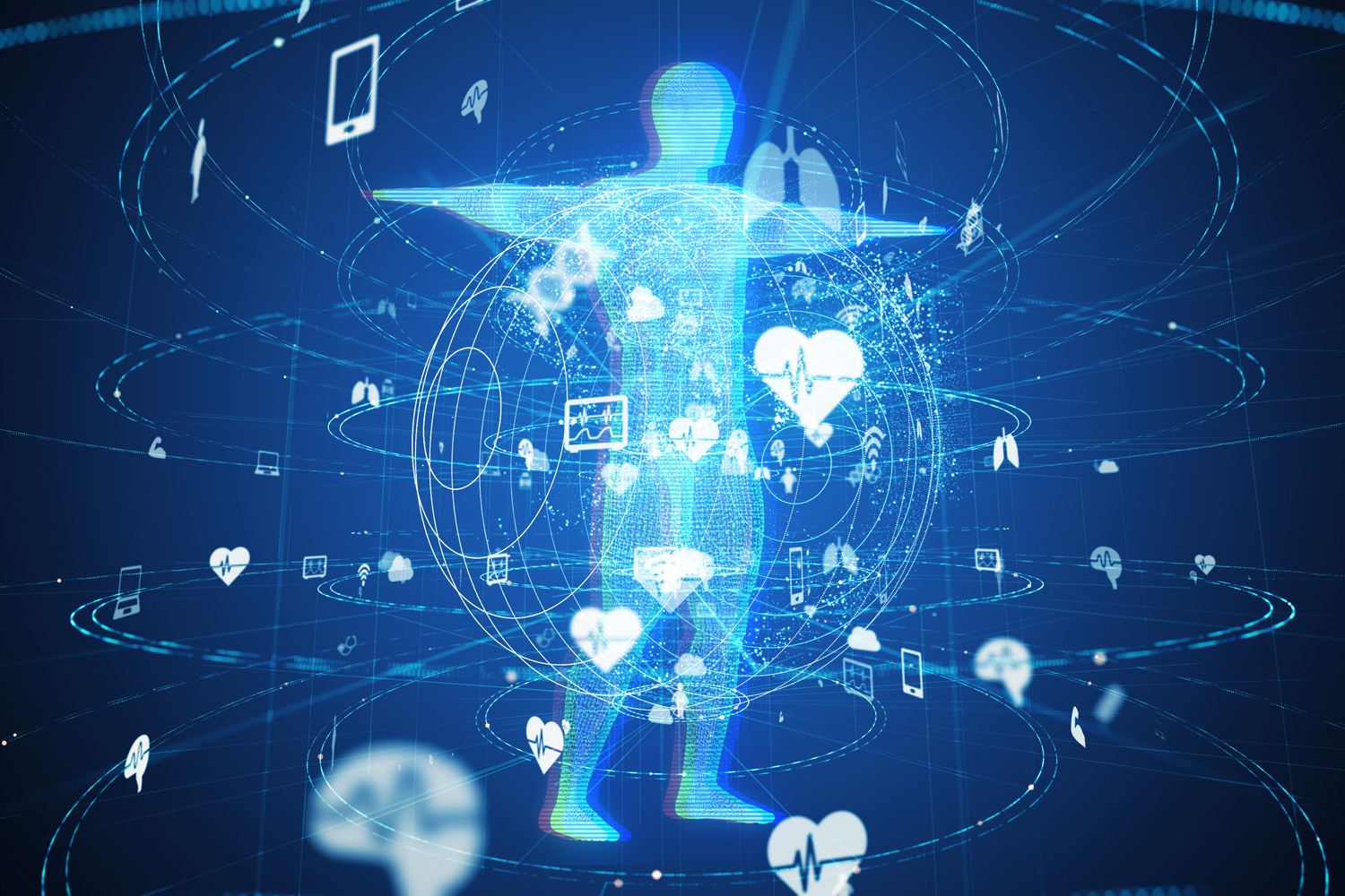
SPECTACULAR NEW COMPUTER TOOLS for managing large amounts of information are moving beyond merely importing, archiving and organizing data. Today, they can generate inferences that allow for more efficient web searching, suggested music and video playlists, highly tailored advertising, and speech and facial recognition. Already available or on the immediate horizon are creative chatbots like ChatGPT and self-driving cars.
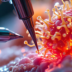
The artificial intelligence (AI) revolution has important implications for all of medicine, and especially for precision cancer care. The first wave of medical AI tools has impacted technologies like mammography and CT scans used in radiographic imaging and microscopy used to examine tissue biopsies and surgical resection specimens.
The AI field may well have been created at the Dartmouth Summer Research Project on Artificial Intelligence, a six-week workshop held in the summer of 1956 at Dartmouth College in Hanover, New Hampshire, that attracted a cadre of forward-thinking mathematicians, engineers, psychologists and others. The attendees tried to imagine what might be needed to create an artificial brain, considering most of the significant domains of the field—domains that remain relevant today.
As computers became faster and large amounts of data became increasingly available, ongoing improvements in algorithmic tools, particularly those involved in statistics-based machine learning, began to deliver impressive achievements. On May 11, 1997, Deep Blue, a chess-playing computer built by IBM, defeated grandmaster Garry Kasparov two games to one, with three draws. On Feb. 16, 2011, IBM Watson defeated Ken Jennings and Brad Rutter, two of the greatest Jeopardy! quiz show champions, earning $77,147 to Jennings’ and Rutter’s $24,000 and $21,600, respectively.
More recently, statistical and machine-learning algorithms, including neural networks and layers of neural networks termed “deep learning,” have allowed increasingly refined pattern recognition and classification. When applied to medical imaging and diagnostic pathology, these types of image analyses can aid in cancer detection and diagnosis, and in determining cancer aggressiveness. Advances in radiographic image acquisition and pathology slide scanning—leading to much larger amounts of data collected—are making AI-aided interpretations increasingly essential.
Historically, radiologists gazed at X-ray pictures and pathologists looked through microscopes. Both used their eyes to resolve features and their brain to recognize patterns from the lower-resolution images obtained through these methods. However, technological advances have increased the resolution and data richness of the images, producing vast amounts of data beyond what the human eye and brain can process, making AI-based assistance invaluable.
The basic strategy used for building AI tools to aid radiology and pathology is to “train” a computer algorithm using a large collection of image files from cases with known outcomes, such as cancer versus noncancer, aggressive cancer versus nonaggressive cancer, and so on, and then to validate the algorithm using a second set of cases. Subsequently, the algorithm can continue to improve as it analyzes more image files.
These approaches will quickly work their way into common clinical practice. In 2021, the Food and Drug Administration released an Artificial Intelligence/Machine Learning Action Plan, anticipating a wave of device applications featuring computer-aided approaches to a wide variety of medical indications. By October 2022, 521 approvals of AI-aided medical devices had been made.
So far, AI has not replaced expert radiologists or pathologists. Rather, the refined classification capabilities have been used to direct the human eye to critical image features. Will this improve precision cancer medicine by evolving into a more highly functioning human-machine interface? As the noted sage Yogi Berra once pointed out, “It’s tough to make predictions, especially about the future.”
Cancer Today magazine is free to cancer patients, survivors and caregivers who live in the U.S. Subscribe here to receive four issues per year.