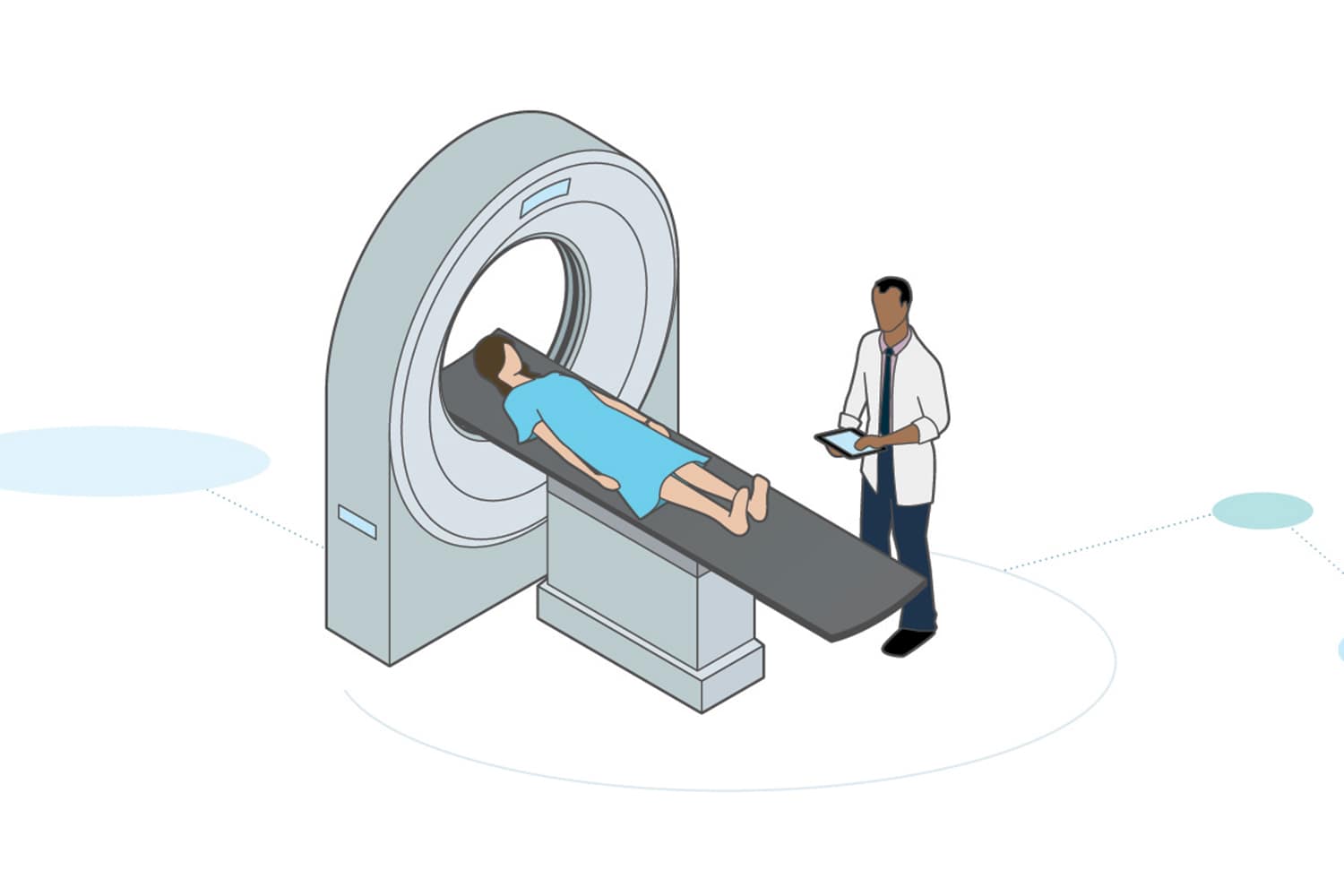
FANCY IMAGING TECHNOLOGIES GIVE PRACTITIONERS detailed pictures of your innards, no knife required. Yet some medical scans involve low doses of radiation, and concern is mounting that their soaring use may nudge up cancer risk. But doctors say prudence, not panic, is the wise response.
A study of six large HMOs published in the June 13 Journal of the American Medical Association found that from 1996 to 2010 the number of ultrasound tests performed per 1,000 enrollees nearly doubled, CT (computed tomography) scans tripled, and MRI (magnetic resonance imaging) exams almost quadrupled. Although ultrasounds and MRIs don’t use radiation, CT scans—like PET (positron emission tomography) scans—deliver more radiation than X-rays.
On average, a person’s annual radiation exposure from CT testing doubled at the six HMOs during the time frame of the study, but about 4 percent of the HMO enrollees received 10 to 20 times the average annual dosage or more in 2010, says Rebecca Smith-Bindman, a radiologist and epidemiologist at the University of California, San Francisco, who led the study.
Potential harms of this radiation exposure include increased cancer risk. In August, a British study linked CT imaging to a small risk of brain tumors. Another study, published in the Archives of Internal Medicine in 2009, estimates that radiation exposure from imaging at current rates could eventually cause 2 percent of future cancers.
According to Bibb Allen Jr., the vice chair of the board of chancellors of the American College of Radiology, the use of sophisticated imaging technologies has grown because they have become integral to patient care. People who receive the most advanced imaging tend to be the sickest, he says, and the tests help sort out complicated medical problems—a need that “is often more pressing than the extremely low risk of cancer many years later.”
Cancer patients in particular often undergo repeat scans, but aggressive CT surveillance of cancer patients in remission might increase the risk of a subsequent tumor, and may not be needed, Smith-Bindman says. Without evidence that frequent scans can detect relapse earlier or improve survival, doing them “doesn’t make sense to me,” she says.
The Children’s Oncology Group, which runs pediatric clinical trials, has been assessing the particular risk posed to kids by medical scans. It recently determined that surveillance CT scans that kids with Hodgkin lymphoma often undergo—typically every four or six months in the first year or two after treatment, and then annually—for up to five years post-treatment “really aren’t necessary” beyond the first year or two, says Cindy Schwartz, a pediatric oncologist at Brown University in Providence, R.I. Schwartz co-authored a July report in the Journal of Clinical Oncology that suggests most relapses occur in the first year, and that detecting relapses after that time does not increase overall survival.
Patients should discuss with their doctors whether a recommended test is really needed, Smith-Bindman says, and if so, how frequently. For example, if your cancer is in remission and the oncologist advises a CT scan every three months, ask whether there’s good evidence the test will benefit you, says Smith-Bindman. If not, see if you could do the scans in three months, then six months after that, then nine months later. More imaging isn’t always better, she says. “I think patients should be a little skeptical,” she adds. “Patients need to be educated consumers, including asking about imaging.”
Cancer Today magazine is free to cancer patients, survivors and caregivers who live in the U.S. Subscribe here to receive four issues per year.