WHEN SANFORD “SANDY” SIMON and his then-12-year-old daughter, Elana, learned in 2008 that her constant belly pains—for years attributed to lactose intolerance, Crohn’s disease or stress—were instead the result of a tumor the size of a large grapefruit in her liver, the next steps weren’t at all clear. The five-year relative survival rate for liver cancer is 21.6%, but Elana’s odds were less straightforward. Her cancer fell into an exceedingly rare and poorly understood category known at the time as the fibrolamellar variant of hepatocellular carcinoma, now fibrolamellar carcinoma (FLC), a type of liver cancer that is more prevalent in adolescents and young adults with otherwise healthy livers.
“The problem [in diagnosing fibrolamellar carcinoma] is that patients have no underlying liver symptoms,” Simon says. “Often patients are told, ‘You’re a teenager; it’s stress.’ Particularly young females are told that for years on end.”
Rare cancers, defined by the National Cancer Institute as those affecting fewer than 40,000 people per year in the U.S., are notoriously difficult to diagnose and treat. With an incidence of 0.02 per 100,000 in the U.S. and only 200 cases each year around the world, FLC remains a diagnosis rarely seen even at major cancer centers. Elana had a one-in-5-million diagnosis, and her doctors and parents had only a handful of small studies to refer to as they determined her treatment course. Researchers didn’t have FLC cell lines or tissue samples to analyze, which meant there were few clues about the biology of the disease.
Both then and now, the primary treatment option for FLC is surgery to remove the liver tumor and surrounding lymph nodes. Elana, whose cancer had not spread to the lymph nodes, had an operation to remove a portion of her liver including the cancerous mass. Simon, who is a cell biologist at Rockefeller University in New York City, worried about the fact that pathologists had identified her liver cancer as FLC based solely on its appearance on scans and not on the molecular characteristics of the tumor cells themselves. The term “fibrolamellar” refers to thick collagen bands surrounding the cancer cells, features that are clearly seen under the microscope. The trouble was, Simon says, any damage to the liver may lead to the formation of bands that look similar, at least on the surface. “When we started, we didn’t know if [her condition] was one disease or many,” Simon says.
Taking the Plunge
FLC, like many rare cancers, wasn’t on the radar as an area to study among most cancer biology researchers, Simon says. Funding agencies and pharmaceutical companies usually prefer to support research with the potential to affect more people. The small number of people with FLC also made it difficult to gather sufficient samples and recruit enough people to do a clinical trial. Although he didn’t study cancer at the time, Simon had a thriving lab and saw a research problem seeking a solution. Elana, with the help of her father, set out to elucidate what was driving FLC.
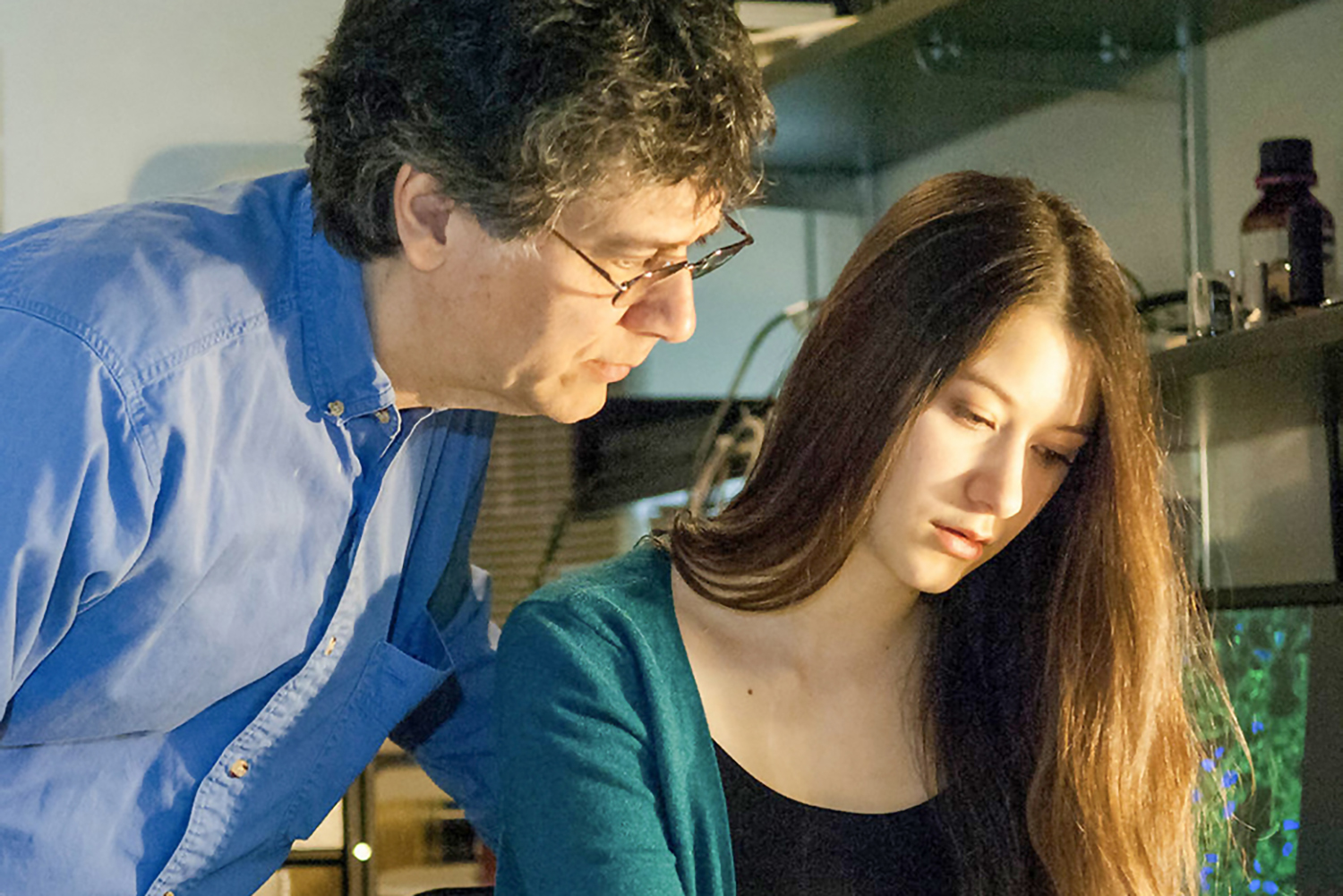
Sanford Simon, a cell biologist at Rockefeller University, and his daughter, Elana, worked to uncover what drove the development of fibrolamellar carcinoma, a rare cancer that affects fewer than 40,000 people per year in the U.S. Photo courtesy of Rockefeller University
In October 2013, Elana and a friend with FLC took to YouTube with a plea for other people with the same cancer to submit tissue samples. The request, unusual at the time, got them their first 15 samples for detailed study in Simon’s lab at Rockefeller University. Not long after, this research led to the discovery of the underlying cause of FLC, findings that were published in Science in February 2014. All the FLC samples shared an unusual loss of DNA, causing two separate genes to run together and produce abnormal “chimeric” RNA molecules. (In Greek mythology, a chimera is a monster with a lion’s head, goat’s body and serpent’s tail. Similarly, chimeric RNA features separate genes combining.)
Every tumor sample had the same chimeric RNA, suggesting the resulting fusion protein might drive FLC and be a promising drug target. Further study revealed that mice engineered to carry the same genetic change and fusion protein got similar liver tumors. In 2016, the Simon family launched the Fibrolamellar Registry, run by patients with FLC and their loved ones to collect health data from medical records and surveys directly from people with FLC. The registry now has 250 participants from 21 countries and makes the data available to other researchers around the world.
“We couldn’t get hospitals to share the information—and we tried—so we set up a nonprofit,” Simon says.
The registry allows patients and family members to contribute their own data even as they move from one cancer center or hospital to the next—which means researchers can track extended outcomes, Simon says. That data has led to a better understanding of FLC over the years. For instance, research generated from the registry found that women diagnosed with FLC generally live longer than men who are diagnosed at the same age and stage. Some FLC tumors also appear to spread more rapidly than others, regardless of their size. The researchers are now attempting to understand what drives these differences.
Facing Logistical Setbacks
Mark Laabs’ experience with cancer at age 28 inspired him to get involved in the rare cancer research community, as well. In 2012, after abruptly losing all vision in his right eye, he learned he had uveal melanoma. The uvea is a tissue layer that includes the iris, or the colored part of the eye. While rare, uveal melanoma is the most common tumor type in the eye, with an incidence rate of 5.1 cases per million people each year. After he received ocular brachytherapy, a type of radiation therapy that uses radioactive seeds on a thin gold plaque that is implanted over the eye, Laabs had no evidence of disease. But uveal melanoma is known to recur and metastasize to other parts of the body in about half the cases.
The only alternative to radiation for uveal melanoma at the time was removing a person’s eye. Laabs, an entrepreneur working in the sustainable energy sector, wondered why doctors didn’t have better treatments for his disease despite progress in treating other forms of melanoma. At 80.9%, the five-year relative survival rate for uveal melanoma in 2013, the year after Laabs’ diagnosis, looked the same as it did in 1973. He soon learned a fundamental cause for this lack of progress was a dearth of tumor samples, which form the building blocks needed to develop cell lines and study cancer in animals. These circumstances were similar for many other rare cancers.
“Scientists didn’t have the basic building blocks to get to the point of asking the interesting scientific questions,” Laabs says.
Mark Laabs launched the Rare Cancer Research Foundation after he was diagnosed with an uncommon cancer of the eye called uveal melanoma. Photo courtesy of Anna Routh Barazin
The primary challenge for researchers, he realized, was a lack of infrastructure to capture rare cancer samples and patients’ associated health data. In 2013, Laabs launched the Rare Cancer Research Foundation (RCRF), a nonprofit dedicated to providing other organizations with the tools they need to engage patients with rare cancers in studies and then make sense of the data. As part of these efforts, Laabs launched pattern.org, a website that allows patients to send tumor tissue samples directly to researchers studying their disease, through the RCRF. Since then, the RCRF has helped people from more than 115 hospitals donate their cancer tissues, leading to cell lines and other models that are enabling the study of rare cancer types.
“No one thought we’d be able to do it,” Laabs says, “but we’ve become one of the largest aggregators of rare cancer samples in the country.”
Gaining Steam
In 2021, the RCRF and the University of Texas MD Anderson Cancer Center in Houston started working together so people with rare cancers could use pattern.org and the RCRF to contribute their samples to MD Anderson for further study. These efforts help to further the reach of MD Anderson’s Rare Tumor Initiative, which met its initial goal to profile tumors from 1,500 people with rare cancers of any type in two years.
MD Anderson has expanded that goal and is now looking to study 10,000 people with a wide range of cancer types. These efforts include careful assessment of the tumor’s DNA, as well as a patient’s inherited DNA and the gut microbiome, says Andy Futreal, chair of Genomic Medicine at MD Anderson. Researchers are also exploring the role of diet, exercise and other lifestyle factors in the risk and progression of cancers. Instead of being an afterthought, patients with rare cancers are “driving the bus” in the effort, Futreal says.
The potential impact of studying rare cancers is surprisingly large. While each rare cancer type affects relatively few individuals, these cancers collectively aren’t rare. One in four people diagnosed with cancer has a tumor that is considered rare. There are currently about 200 rare cancer types, affecting more than 400,000 people in the U.S. each year.
“We see a lot of rare cancer patients at MD Anderson,” Futreal says, “and we have a moral obligation to pay attention to rare cancers [just] as we do with more common ones.”
Rare Discoveries
Breast pathologist and physician-scientist Fresia Pareja and research scientist Britta Weigelt study rare breast and gynecologic cancers at Memorial Sloan Kettering Cancer Center in New York City. They note fundamental discoveries in rare cancer have potential implications for other cancers, too.
“[The study of rare cancers] is an opportunity for the discovery of novel cancer genes,” Pareja says.
Discovering just one single genetic change in a cancer, even if it is rare, can open the door to understanding more common cancers that can share similar features. In fact, the ability to make such fundamental discoveries through studies of rare cancers has a long history. For instance, building on more than a decade of observations, scientists found the first tumor suppressor gene ever described in a rare eye cancer called retinoblastoma in 1986.
There is still more to learn, researchers note. In 2018, a study published in Nature Communications by Pareja, Weigelt and colleagues found that two genes are mutated and appear to act as tumor suppressors in granular cell tumors, a rare tumor type. The genes, called ATP6AP1 and ATP6AP2, are known to control the pH, or acidity, inside cellular compartments. In addition to highlighting unknown tumor suppressors, the findings offered genetic evidence that a breakdown in pH control might play a role in some cancers.
Get the information that will help you understand your diagnosis and treatment options as someone with a rare cancer.
When you or a loved one has a rare cancer diagnosis, it’s always a good idea to gather as much information as you can about the cancer type and ask doctors plenty of questions, including:
- Have you diagnosed and treated this cancer type before?
- Should I have my cancer genetically tested or sequenced?
- Can I donate my tissue for rare cancer research?
- Is there an organization, registry or social media group for my cancer type?
- Where can I find out about clinical trials?
If you aren’t completely satisfied with the answers you’re getting or need more perspective, be sure to seek out a second opinion at a cancer center where doctors see many people with rare cancers, if possible.
The discovery of underlying genetic alterations in rare tumors may help refine cancer classification systems, Pareja says, which could result in improvements in the diagnosis of rare tumors. For instance, scientists from Weigelt’s lab discovered a unique genetic change in a rare and noncancerous ovarian tumor called sclerosing stromal tumor, which can be misdiagnosed as ovarian cancer. Their discovery may lay the foundation for testing to ensure that people with these tumors are not misdiagnosed or overtreated.
“Having the correct diagnosis is of great importance,” Pareja says. “If you have a marker that can help you say [for certain] it is this [rare] tumor type, that’s extremely helpful.”
Optimism for the Future
While clinical trials studying cancers that affect very few people are more challenging to conduct, Simon says work in his daughter’s rare liver cancer has led to promising treatment leads. He expects his work will lead to a clinical trial that could enroll people with FLC as soon as 2024. Similarly, Laabs says there is now a better understanding of the genetic alterations underlying uveal melanoma, but when it comes to treatments that might extend survival or cure the disease, “it’s a work in progress.”
After nine years in remission, Laabs was diagnosed in late 2020 with a metastatic recurrence of his rare melanoma in the membrane that lines his lungs and chest. Despite the rarity of his diagnosis and limited treatment options, he had his primary and metastatic cancer’s DNA sequenced and has enrolled in clinical trials aimed at producing custom-made, experimental vaccines and cell therapies based on his tumor’s molecular profile, both geared to ramp up an immune response against his specific cancer. While it’s not clear if that has helped him beat the odds so far, Laabs is generally optimistic about the future for people with rare cancers, including uveal melanoma, even if there are no “silver bullets.”
These efforts by advocacy organizations join federal initiatives as well, which aim to increase understanding of rare cancers. For instance, the Food and Drug Administration Oncology Center of Excellence, authorized in 2016 by the 21st Century Cures Act, includes the Rare Cancers Program, which is dedicated to engaging stakeholders in developing and providing expedited review of promising new treatments for rare cancers. In addition, the National Cancer Institute launched an effort known as the My Pediatric and Adult Rare Tumor Network, enlisting scientists, patients, advocates and doctors to work together to help find treatments for rare cancers.
“We’re generating way more data now as a cancer research community overall, and the usefulness of that data is skyrocketing,” Laabs says. “The pace of innovation in cancer treatment is by far the fastest it has ever been since I was diagnosed. Treatments today are meaningfully different from one year ago. As a patient, I draw a lot of excitement about that, and it basically means my job as a patient with a cancer for which we have not found a cure is to stick around while this awesome innovation is happening, and I think I can, and I think a lot of other patients can, who not long ago may have had fewer options.”
Cancer Today magazine is free to cancer patients, survivors and caregivers who live in the U.S. Subscribe here to receive four issues per year.